Skip to content

Illustration of ankle injuryAnkle Injuries

The ankle is the most injured peripheral joint in the body. Ankle injury ranges from 15% to as high as 59% of all injuries in some sports. The sports that most commonly produce high ankle injury rates in their participating athletes include: basketball, netball, and the various codes of football. Thus those sports and other activities that involve running, balance and quick stop-start movements appear to be of higher risk. There is a two-fold increased chance of ankle injury in those that have previously sustained an ankle injury compared to those that have not.

Injury

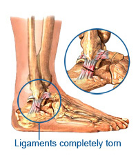
The mechanisms of ankle ligament injury are varied but are commonly due to inversion coupled with plantar flexion. Thus, lateral ankle ligament injury is most common and is often associated with peroneal tendon strain or rupture. Lateral ankle ligament injury account for in excess of 40% of all ankle injuries. Medial ankle ligament injuries are less common. This is due to the strength of the deltoid ligament and structure of the ankle joint itself. However, medial injury may occur with excessive pronation, eversion and dorsiflexion, or pronation and external rotation. These movements may also induce sprains to the ankle syndesmosis, such injury only occurs in 10% of ankle injuries. If there is a history of compressive forces to the mortise, osteochondral damage needs to be ruled out. Ligament injury is graded depending on the degree of damage. Grade 1 injuries cause stretching of the ligament without macroscopic tear, and the joint is considered stable on testing. Grade 2 injuries consist of partial macroscopic tearing with mild to moderate instability. Moderate swelling and tenderness are present and the functional ability is compromised. The Grade 3 sprain involves complete ligament rupture associated with marked swelling, ecchymosis and instability. Differential diagnosis of lateral ankle injury: Pain that persists following an acute lateral ankle sprain should alert the clinician to the possibility of other injury concomitant with the ligament injury. The differential list should include several possibilities not limited to: chronic instability, early degenerative joint disease, loose bodies, osteochondral lesions, occult fractures, intra-articular meniscoid lesions and peroneal tendon injury (including tendinitis, rupture and subluxation).

Assessment

A detailed history is needed to determine mechanism of injury. The patient may often describe a ‘crack’ or popping sound. This, along with the level of swelling is a good indictor of severity. Often with a sprain, the patient is able to weight-bear immediately after the injury, and often report continuing activity. Site of pain gives a good indication of the tissues damaged. Special tests such as anterior draw (moving talus anterior), talar tilt (moving the talus laterally and medially) and proprioception are used to assess the degree of ankle integrity. The Ottawa ankle rules on X-Ray imaging are used to determine if an X-Ray is required.

Treatment Types

Treatment regimes for lateral ankle sprain are indeed quite varied. The injury type often dictates the form of the treatment, but in many cases, several approaches have been used for the same injury. The most commonly used forms of treatments are mobilisation, immobilisation, and surgery. Mobilisation:

Early mobilisation (pain limited weight bearing activity) is by far the most common approach in contemporary sports medicine. Mobilisation is a generic term for treatments where the ankle joint is left relatively free to move in the first few days to weeks following trauma. As such, there are numerous variations and modifications on the general theme. It is seen as a low cost, high-result therapy that has been shown on multiple occasions to be more effective in getting injured ankles functioning quicker than other treatment forms including rest and immobilisation. It is also noted that these therapies are safe and complication free.

Immobilization

In cases where immobilisation is used (generally grade 2 or 3 strains), the ankle is prevented from moving as soon as the diagnosis is made. Immobilization is achieved most commonly through a plaster cast, although fibreglass and air casts are also being used. The period of immobilisation ranges between 10 days (1) to 4 or 6 weeks. Following this period rehabilitation phases 2 and 3 as seen below are usually undertaken. Studies performed on the comparisons of mobilisation and immobilisation have noted that although restoration of ankle function is quickest with mobilisation, they also report that in follow up examinations, the long term results are similar between groups. To date, the literature seems to suggests that immobilisation is best performed in those patients who suffer chronic lateral instability or are professional athletes, and then only in conjunction with surgery. Although one study has provided evidence to suggest that the treatment of athletes should incorporate functional / mobilisation techniques.

Surgery

Surgery is indicated in the case of instability and joint decompression secondary to loose or foreign bodies (meniscoid lesions, impingement and talar dome osteochondral injuries).

Rehabilitation

The program goals are to minimize the effusion, normalize gait, normalize pain-free range of motion, prevent muscular atrophy, maintain proprioception, and maintain cardiovascular fitness. Most methods involve a 3-phase treatment regime started as soon as the injured ankle is seen. Phase One

The first phase utilizes standard first aid (PRICER) treatment. Ankle rest is usually performed with the use of crutches. Initially the patient is non-weight bearing with progression to weight bearing as the pain allows. Soft tissue therapy to resolve spasm of the gastrocnemius-soleus complex can also commence. The acute phase generally lasts for a period of 1 to 3 days, depending on the grade of injury.

Phase Two

The second phase immediately follows the first phase with the time frame varying for each grade of injury. For a grade one injury, the second phase would generally start on day two and last two to four days. In a grade two injury it may start on day two to three and last eight to twelve days. With a grade three injury, the second phase may start in six to ten days and last three to four weeks. The second phase consists of general ankle muscle strengthening, notably of the dorsiflexor and peroneal groups, and stretching the achilles tendon. Exercises such as toe writing (where the alphabet is scribed by the foot in the air), and plantar and dorsiflexion movements against resistance (eg rubber tubing or bands) may be performed during this phase. Soft tissue therapy to facilitate proper scar tissue formation and maintainance of joint function is continued.

Phase Three

The third phase involves conditioning and proprioceptive training. Materials commonly used include wobble boards, mini tramps and rocker boards, which are used in conjunction with a regime of increasing functional activities (ie progressing from brisk walking, to running, etc, and ultimately to jumping, hopping and cutting). The functional activities progress through to sport-specific exercises for the patient. This phase follows the second phase and starts when the patient has 80% strength returned to the injured site.

For further information on Ankle Injuries and Professional Treatment available at Synergy Healthcare contact us today. CONTACT US


Ankle Injuries Sylvania Waters | Synergy Healthcare